The neuromuscular junction (NMJ) is a specialized synapse or junction between a motor neuron and a muscle fiber (muscle cell). It is a crucial component of the neuromuscular system, where nerve signals are transmitted to muscles, ultimately leading to muscle contraction. Here’s a detailed note on the structure and function of the neuromuscular junction:
Structure of the Neuromuscular Junction:
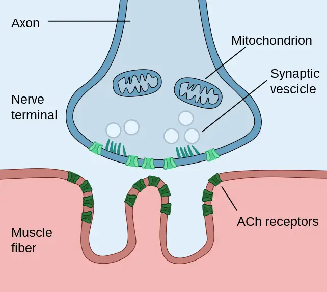
1. Motor Neuron Terminal: The process begins with a motor neuron, which is a nerve cell that carries signals from the central nervous system (CNS) to muscles. The motor neuron terminates at the neuromuscular junction.
2. Synaptic End Bulb: The distal end of the motor neuron axon forms a bulbous structure known as the synaptic end bulb or synaptic terminal. Within the synaptic end bulb are numerous synaptic vesicles containing the neurotransmitter acetylcholine (ACh).

3. Synaptic Cleft: Separating the synaptic end bulb of the motor neuron from the muscle fiber is a narrow, fluid-filled gap called the synaptic cleft.
4. Motor End Plate: The motor end plate is the specialized region of the muscle fiber’s sarcolemma (cell membrane) facing the synaptic end bulb. It contains numerous ACh receptors.
Function of the Neuromuscular Junction:
1. Nerve Impulse Transmission:
– The process begins when an action potential (nerve impulse) reaches the synaptic end bulb of the motor neuron. This action potential is generated in response to a signal from the central nervous system, such as a voluntary muscle contraction command.
– Depolarizing the motor neuron’s membrane causes voltage-gated calcium channels in the synaptic end bulb to open. Calcium ions (Ca2+) enter the synaptic end bulb from the extracellular fluid.
– The influx of calcium ions triggers the fusion of synaptic vesicles with the motor neuron’s membrane, releasing acetylcholine (ACh) into the synaptic cleft via exocytosis.
2. ACh Binding to Receptors:
Acetylcholine diffuses across the synaptic cleft and binds to ACh receptors on the motor end plate of the muscle fiber. These receptors are ligand-gated ion channels.
3. Depolarization of the Sarcolemma:
– Binding of ACh to its receptors initiates the opening of sodium (Na+) channels in the sarcolemma of the muscle fiber. Sodium ions rush into the muscle fiber, causing a local depolarization of the membrane.
– This local depolarization, known as an end-plate potential (EPP), spreads throughout the muscle fiber’s sarcolemma.
4. Propagation of Muscle Action Potential:
– If the EPP is sufficient to reach the threshold for muscle action potential generation, it will trigger the opening of voltage-gated sodium channels along the sarcolemma.
– The muscle action potential travels along the sarcolemma and into the T-tubules (transverse tubules), which are invaginations of the sarcolemma, allowing the action potential to reach the interior of the muscle fiber quickly.
5. Calcium Release from Sarcoplasmic Reticulum:
– The muscle action potential in the T-tubules triggers the release of calcium ions (Ca2+) from the sarcoplasmic reticulum (SR), a specialized organelle within the muscle fiber.
6. Muscle Contraction:
– Calcium ions bind to troponin, a regulatory protein on the thin filaments within the muscle fiber’s sarcomeres.
– This binding causes a conformational change in troponin and allows the myosin heads (thick filaments) to interact with actin (thin filaments), initiating the sliding filament mechanism and muscle contraction.
7. Termination of Muscle Contraction:
– Muscle contraction continues as long as action potentials are generated, and calcium ions are available.
– Muscle contraction terminates when the action potential ceases, and the sarcoplasmic reticulum actively pumps calcium ions back, inducing muscle relaxation.
The neuromuscular junction is a critical site for transmitting nerve signals to muscle fibers, allowing precise control over muscle contractions. Dysfunction or disorders affecting this junction can lead to muscle weakness, paralysis, and neuromuscular diseases, making it an essential area of study in neurophysiology and clinical medicine.


